Nerve Spine Chart
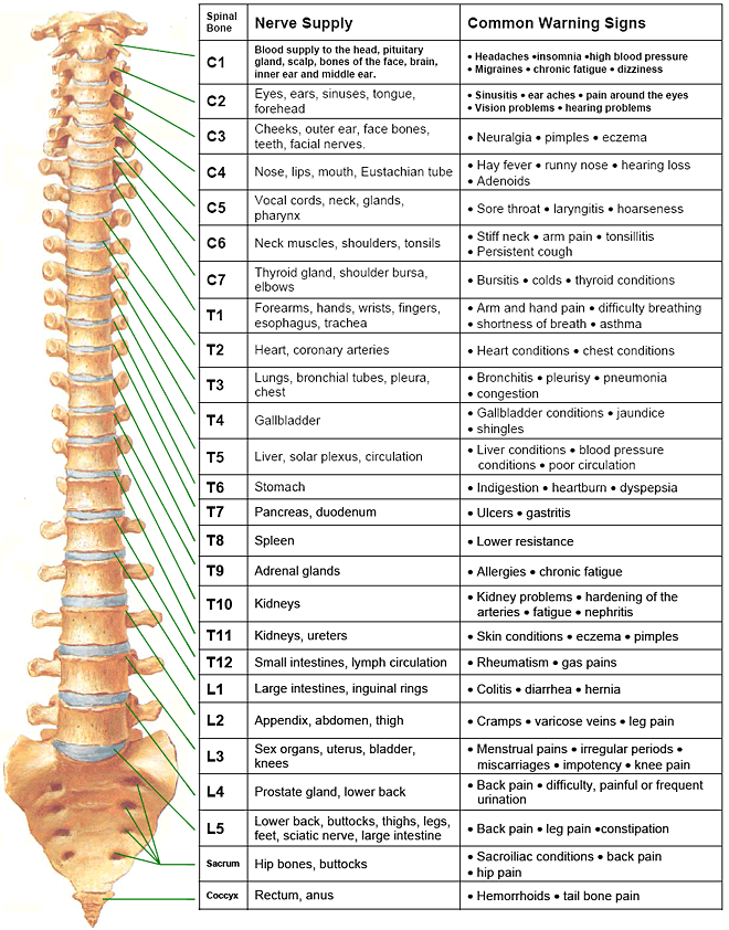
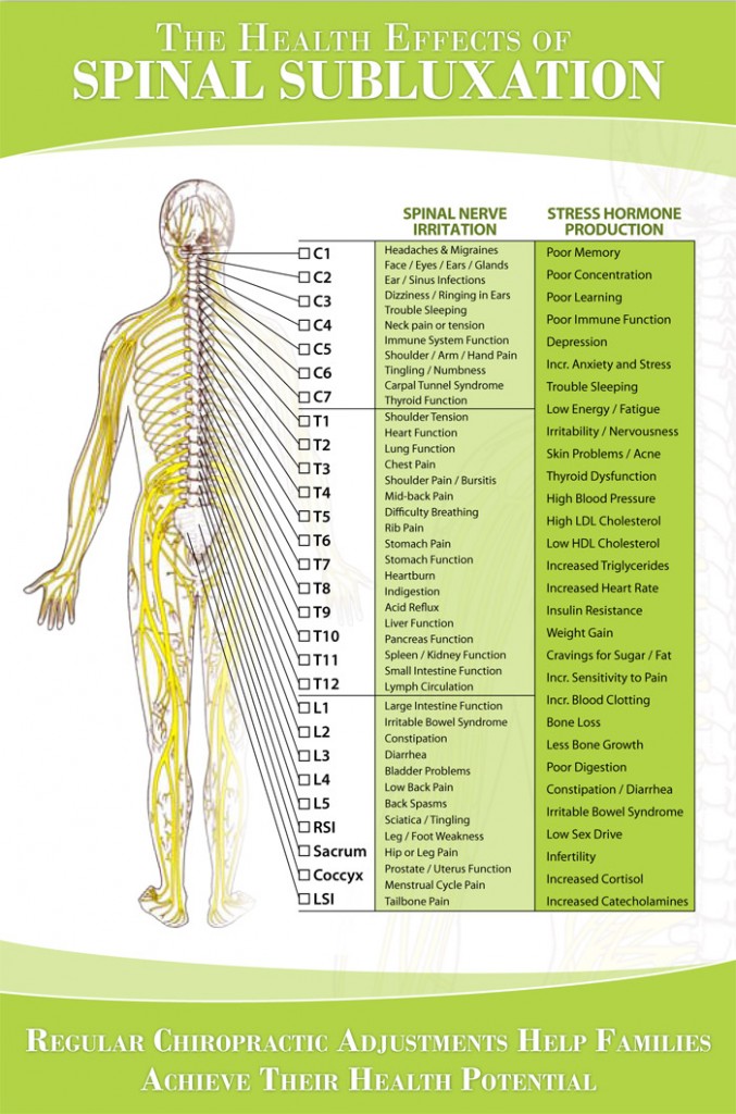
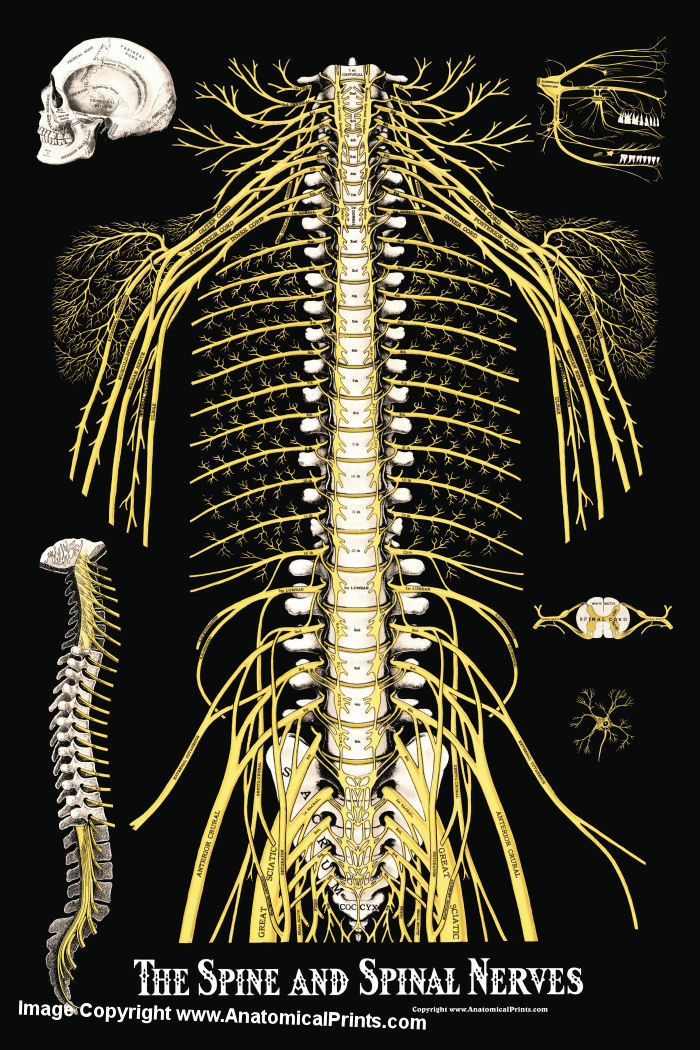
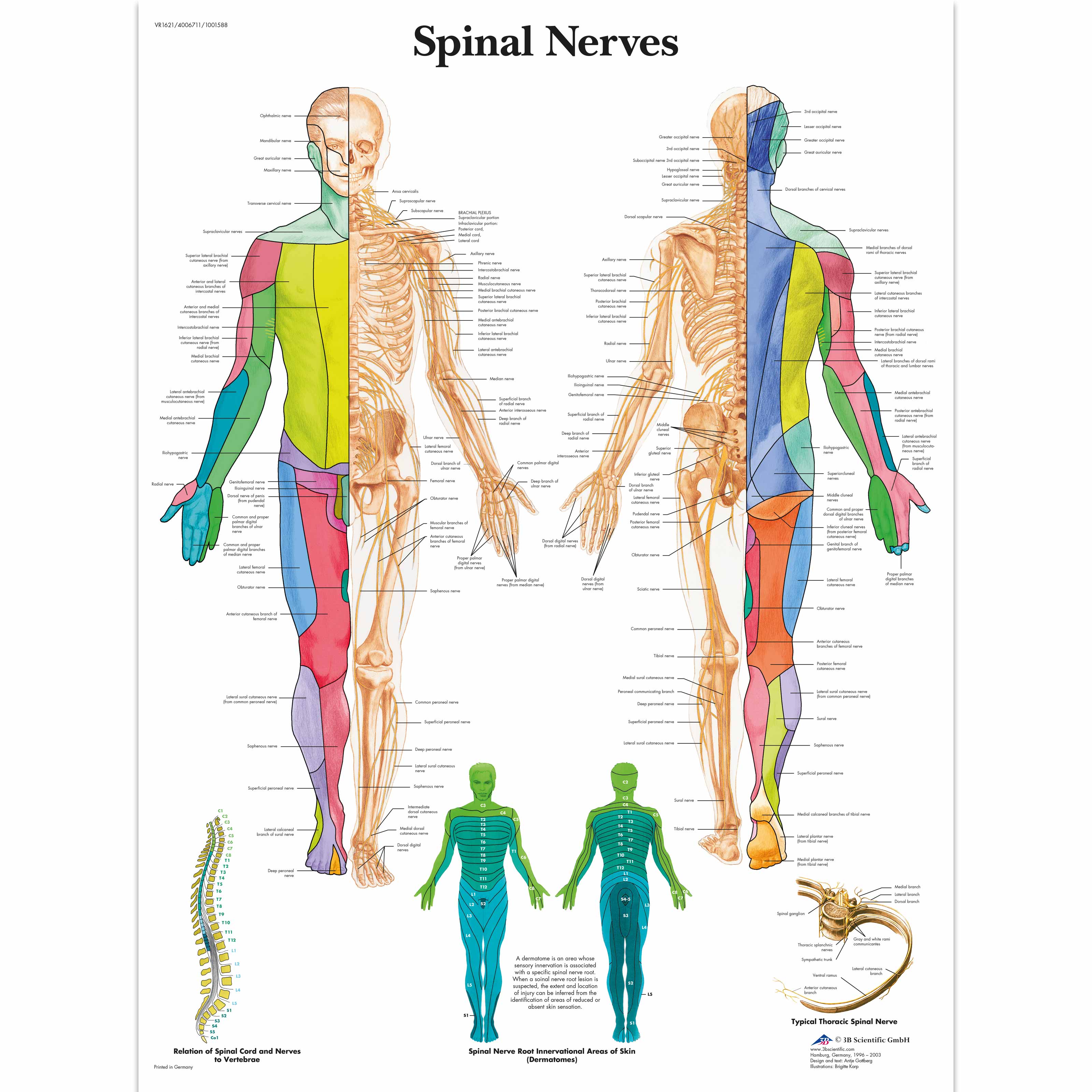
Nerve Spine Chart - Web learn how spinal nerve roots function, and the potential symptoms of spinal nerve compression and pain in the neck and lower back. Web chiropractic spinal nerve chart is used by chiropractors to show nerve connections at each segmental level. Click to view larger image. These nerves also control movements of the hip and knee muscles. The nervous system consists of the brain, spinal cord, sensory organs, and all of the nerves that connect these organs with the rest of the body. New 3d rotate and zoom. Each spinal nerve originates from two roots: Spinal nerves emerge from the spinal cord and reorganize through plexuses, which then give rise to systemic nerves. For the most part, the spinal nerves exit the vertebral canal through the intervertebral foramen below their corresponding vertebra. This root has a swelling called the dorsal root ganglion, which contains the cell bodies of sensory neurons. L2, l3, and l4 spinal nerves provide sensation to the front part of the thigh and inner side of the lower leg. Web these relay motor (movement), sensory (sensation), and autonomic (involuntary functions) signals between the spinal cord and other parts of the body. Web a dermatome is a distinct area of your skin defined by its connection to one of 30 spinal nerves. We’ll explore more about both your spinal nerves and dermatomes, including a chart showing each. Web spinal nerves are all mixed nerves with both sensory and motor fibers. Web there are 31 bilateral pairs of spinal nerves, named from the vertebra they correspond to. Web learn how spinal nerve roots function, and the potential symptoms of spinal nerve compression and pain in the neck and lower back. Together, these organs are responsible for the control of the body and communication among its parts. Each spinal nerve originates from two roots: Spinal nerves can be impacted by a variety of medical conditions, resulting in pain, weakness, or decreased sensation. Carries sensory (afferent) information to the spinal cord. Web below is a chart that outlines the main functions of each of the spine nerve roots: This root has a swelling called the dorsal root ganglion, which contains the cell bodies of sensory neurons. An essential chiropractic nerve function chart. Spinal nerves emerge from the spinal cord and reorganize through plexuses,. It is important to mention that after the spinal nerves exit from the spine, they join together to form four paired clusters of. Spinal nerves can be impacted by a variety of medical conditions, resulting in pain, weakness, or decreased sensation. Each spinal nerve originates from two roots: Carries sensory (afferent) information to the spinal cord. Web l1 spinal nerve. The nervous system consists of the brain, spinal cord, sensory organs, and all of the nerves that connect these organs with the rest of the body. Spinal nerves can be impacted by a variety of medical conditions, resulting in pain, weakness, or decreased sensation. This root has a swelling called the dorsal root ganglion, which contains the cell bodies of. An essential chiropractic nerve function chart. These nerves also control movements of the hip and knee muscles. Together, these organs are responsible for the control of the body and communication among its parts. This root has a swelling called the dorsal root ganglion, which contains the cell bodies of sensory neurons. It is important to mention that after the spinal. Together, these organs are responsible for the control of the body and communication among its parts. Web there are 31 bilateral pairs of spinal nerves, named from the vertebra they correspond to. This root has a swelling called the dorsal root ganglion, which contains the cell bodies of sensory neurons. Spinal nerves emerge from the spinal cord and reorganize through. Each spinal nerve originates from two roots: This root has a swelling called the dorsal root ganglion, which contains the cell bodies of sensory neurons. Web there are 31 bilateral pairs of spinal nerves, named from the vertebra they correspond to. These nerves also control movements of the hip and knee muscles. L2, l3, and l4 spinal nerves provide sensation. L2, l3, and l4 spinal nerves provide sensation to the front part of the thigh and inner side of the lower leg. Web l1 spinal nerve provides sensation to the groin and genital regions and may contribute to the movement of the hip muscles. For the most part, the spinal nerves exit the vertebral canal through the intervertebral foramen below. Web these relay motor (movement), sensory (sensation), and autonomic (involuntary functions) signals between the spinal cord and other parts of the body. New 3d rotate and zoom. Spinal nerves emerge from the spinal cord and reorganize through plexuses, which then give rise to systemic nerves. The nervous system consists of the brain, spinal cord, sensory organs, and all of the. Together, these organs are responsible for the control of the body and communication among its parts. This root has a swelling called the dorsal root ganglion, which contains the cell bodies of sensory neurons. It is important to mention that after the spinal nerves exit from the spine, they join together to form four paired clusters of. Web spinal nerves. New 3d rotate and zoom. We’ll explore more about both your spinal nerves and dermatomes, including a chart showing each. Web l1 spinal nerve provides sensation to the groin and genital regions and may contribute to the movement of the hip muscles. Web these relay motor (movement), sensory (sensation), and autonomic (involuntary functions) signals between the spinal cord and other. An essential chiropractic nerve function chart. For the most part, the spinal nerves exit the vertebral canal through the intervertebral foramen below their corresponding vertebra. Web l1 spinal nerve provides sensation to the groin and genital regions and may contribute to the movement of the hip muscles. Spinal nerves can be impacted by a variety of medical conditions, resulting in pain, weakness, or decreased sensation. We’ll explore more about both your spinal nerves and dermatomes, including a chart showing each. Spinal nerves emerge from the spinal cord and reorganize through plexuses, which then give rise to systemic nerves. L2, l3, and l4 spinal nerves provide sensation to the front part of the thigh and inner side of the lower leg. This root has a swelling called the dorsal root ganglion, which contains the cell bodies of sensory neurons. Together, these organs are responsible for the control of the body and communication among its parts. The nervous system consists of the brain, spinal cord, sensory organs, and all of the nerves that connect these organs with the rest of the body. Web learn how spinal nerve roots function, and the potential symptoms of spinal nerve compression and pain in the neck and lower back. Web these relay motor (movement), sensory (sensation), and autonomic (involuntary functions) signals between the spinal cord and other parts of the body. These nerves also control movements of the hip and knee muscles. Web chiropractic spinal nerve chart is used by chiropractors to show nerve connections at each segmental level. Each spinal nerve originates from two roots: Web spinal nerves are all mixed nerves with both sensory and motor fibers.
Lumbar Spinal Nerve Chart
Printable Spinal Nerve Chart
Nerve Chart Hunter Chiropractic Wellness Centre

Spinal Nerve Function Anatomical Chart Anatomy Models and Anatomical
The Spine and Spinal Nerves Poster Clinical Charts and Supplies
Anatomical Charts and Posters Anatomy Charts Spinal Nerves
![Free Printable Spinal Nerve Charts [Function & Diagram] PDF](https://www.typecalendar.com/wp-content/uploads/2023/08/Spinal-Nerve-Chart-Example-PDF.jpg)
Free Printable Spinal Nerve Charts [Function & Diagram] PDF

Spinal Nerve Chart

Printable Spinal Nerve Chart Printable World Holiday

Printable Spinal Nerve Chart Printable World Holiday
Carries Sensory (Afferent) Information To The Spinal Cord.
Web Below Is A Chart That Outlines The Main Functions Of Each Of The Spine Nerve Roots:
It Is Important To Mention That After The Spinal Nerves Exit From The Spine, They Join Together To Form Four Paired Clusters Of.
Thoracic Spinal Nerves Are Not Part Of Any Plexus, But Give Rise To The Intercostal Nerves Directly.
Related Post: